Filter Categories
Latest
Customer stories
Parity News
White papers & articles
Product Updates

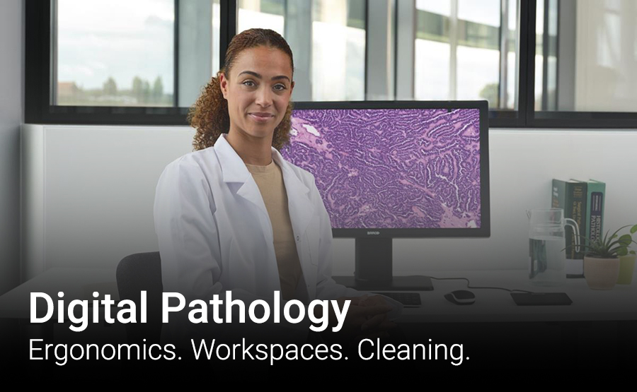
In this article we will explain what Digital Pathology is, why it’s needed and the landscape of Digital Pathology in the NHS. We’ll explore the benefits of it, but also the challenges of introducing new technology and how to ensure that areas are not congested, cleaning protocols are ensured and ergonomics are considered.
“Pathology facilities should be designed for easy, frequent and thorough cleaning” – NHS England.
Digital pathology is an emerging technology in pathology, in which a scanner converts glass tissue slides into digital slides that can be viewed and analysed on a display with the help of viewing software, after which they are stored digitally.
Source – Barco Healthcare Glossary.
More pathology tests are needed each year, driven by an aging population, increasing cancer rates and the growth of precision medicine, and resulting in pathologist workload significantly growing every year. Some regions, such as a number of countries in Western Europe, are well into the digital transformation and realizing meaningful patient care advantages every day, others are following at an accelerating pace.
In order to partly compensate the looming shortage of pathologists worldwide, digital pathology offers opportunities through the application of new and emerging technologies.
Digital pathology can facilitate faster, more efficient diagnoses and prognoses, more flexible collaboration and high-quality case documentation. Online storage of digital slides also opens doors for deep learning and artificial intelligence applications.
In order to partly compensate the looming shortage of pathologists worldwide, digital pathology offers opportunities through the application of new and emerging technologies.
With a lot of digital information amassed, big data and deep learning mechanisms can contribute to the development of computer- or even AI-assisted diagnostic tools. These tools can serve as ‘second pairs of eyes’, automating and enhancing the recognition of specific biomarkers in tissue sections, but they can also help analysing large sets of slides, classifying them and providing overarching insights about specific anomalies, for instance. In the long term, this can lead to more accurate predictions of diseases.
Source – Barco Healthcare.

Efficiency
With digital slides accessible online, slide handling and archiving offer many possibilities, almost all of which contribute to faster turnaround times and more efficient workflows. A digitized field makes it possible to expand workflow capacity and speed up second opinions, because they can be done remotely and slides don’t need to be shipped physically for consultations.
Case documentation
Digital files also make digital annotations possible, which can be shown or hidden at the pathologist’s will. These annotations can be side remarks about the case or specific notations on the image itself, highlighting a specific area for example. They are linked to the digital file but don’t change the integrity of the source image itself.
Telepathology
Analog telepathology can be considered the predecessor of modern whole slide imaging systems, involving the live remote control of a microscope. Remote parties can see the slide as viewed through a microscope and join in its discussion.
Digital pathology makes remote consultation easier and offers more possibilities, as the same file can be opened and viewed in multiple places at any time. With the right displays and viewing software, digital slides can be viewed in exactly the same way by remote participants. Work can be outsourced to specialized facilities if necessary, and a digital workflow also clears the way for the pathology staff to work at home or elsewhere.
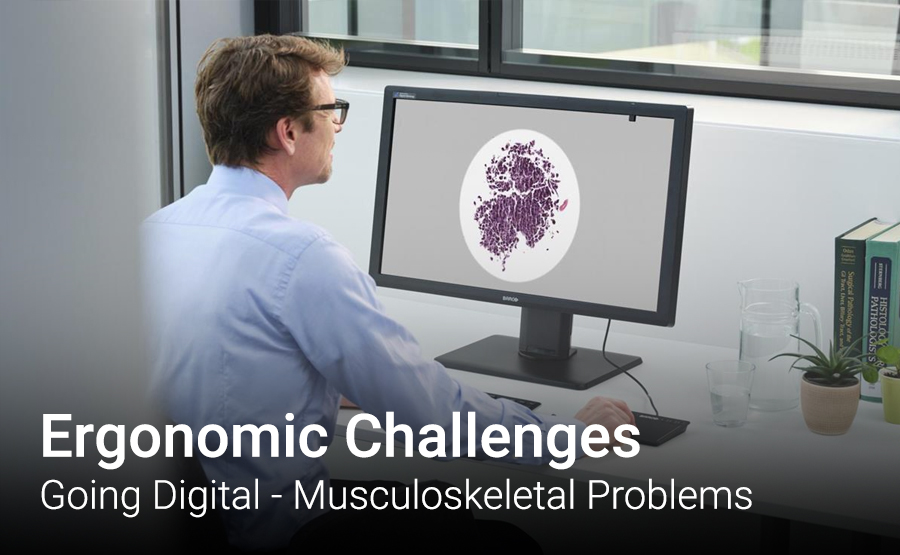
We have seen the NHS using Radiology as an example when rolling out Digital Pathology. There are similarities to when ‘PACs’ was introduced, and Digital Pathology.
The benefits were significant, however they also had unforeseen challenges as Reporting rooms were not set up for ergonomics. This can lead to long term problems such as an increase in musculoskeletal disorders.
“The transition from a film-based to a filmless soft-copy picture archiving and communication system (PACS)-based environment has resulted in improved work flow as well as increased productivity, diagnostic accuracy, and job satisfaction…. However, the importance of optimising workplace ergonomics has received little attention. Factors such as the position of the work chair, workstation table, keyboard, mouse, and monitors, along with monitor refresh rates and ambient room lighting, have become secondary considerations. Paying close attention to the basics of workplace ergonomics can go a long way in increasing productivity and reducing fatigue, thus allowing full realisation of the potential benefits of a PACS. Optimisation of workplace ergonomics should be considered in the basic design of any modern radiology suite.”
Source: Importance and Effects of Altered Workplace Ergonomics in Modern Radiology Suites.
In a study of 148 Radiology Consultants and Trainees, across 10 Hospital sites – it was found that 38% reported radiology-associated occupational injury. Lower back discomfort was the commonest radiology associated musculoskeletal symptom (41%).
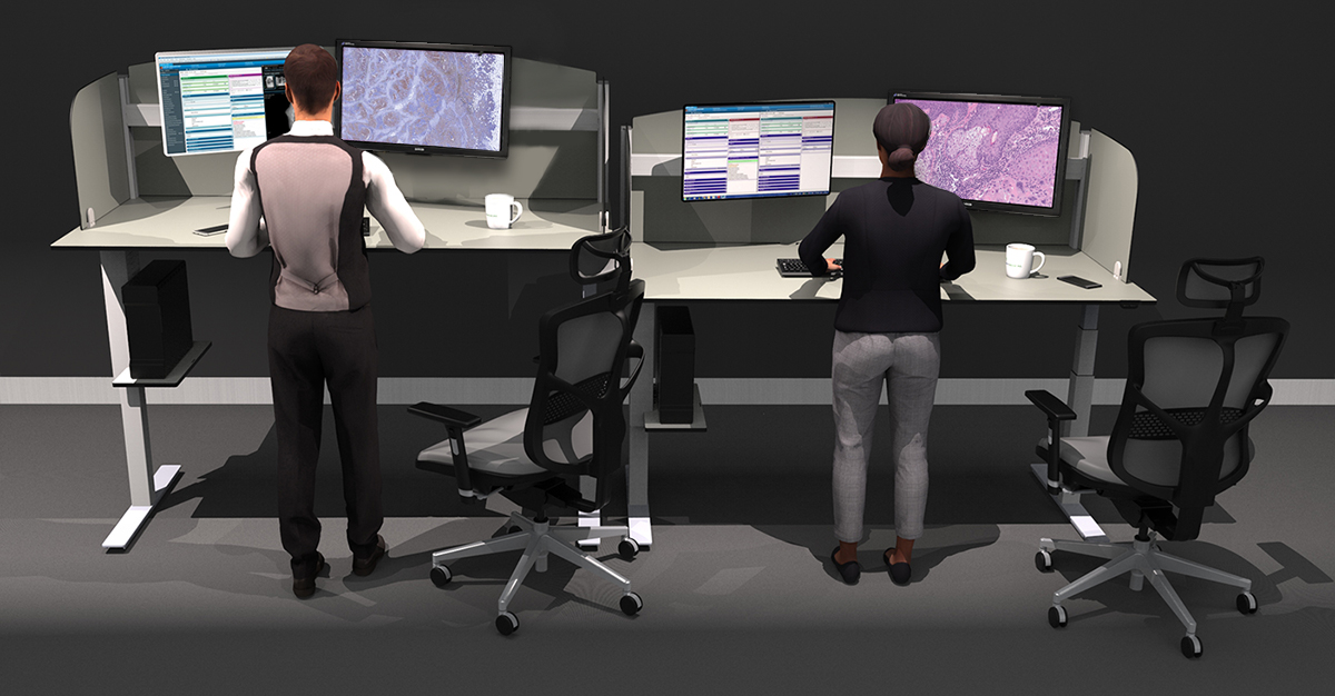
Digital Pathology will naturally spend longer sat in a chair, staring at a display. Learning from Radiology, and using the data above – one solution to this was the introduction of sit-stand desks.
With 40% of all sickness absence in the NHS is due to musculoskeletal disorders. A British Medical Journal study showed that NHS staff who used sit-stand workstations resulted in fewer musculoskeletal problems. Alternating between sitting and standing every 30 minutes has been shown to reduce musculoskeletal symptoms by up to 32%.
Sit-stand working can make long hours spent reporting more comfortable and improves health, resulting in considerable savings by reducing absence from work caused by poor ergonomics.
Our Energise Sit-Stand Desk has been the go-to solution to alleviate those challenges seen in Radiology, and now it has been the most requested solution we have seen from Pathologists/Project Mangers looking at Digital Pathology.
With that in mind, we have designed a new ‘Sit-Stand Digital Pathology Workstation’, which is an fully configured Workstation designed for Pathologist workflow. Learn more here.
As well as the sit-stand benefits, this solution also can be provided/configured with:
Even with a sit-stand desk, naturally a Pathologist will still sit for long durations. This is because it’s recommended that staff alternate between both, rather than completely standing or completely sitting.
We can provide our Reporting Chair which is an ergonomic chair that is proven to increase performance and is perfectly suited to support Pathologists who spend long hours looking at screens. It’s designed to support every part of you, so that you can stay healthy, comfy and completely focused on your work.
An estimated 70 to 80% (NHS Review Of Pathology Services) of all healthcare diagnoses and decisions are directly influenced by Pathology test results. Modern equipment helps with the speed and reliability of tests performed, and means a better service for patients.
An increase in this equipment can create congested areas, as well as challenges when accessing the new equipment safely, ergonomically, and being compliant with cleaning protocols.
By clearing benches and Wall Mounting equipment – you make everything easier to clean and a better working environment for all.
We would recommend an audit of the working space, and we can help you with the following:
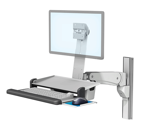
An increase in technology within Pathology can lead to congested areas and cluttered benches. By clearing your benches, you free up space to work while also creating easy to clean worksurfaces. But how can you keep them clear? Wall Mounts!
The go-to solution is to move technology onto medical-grade Wall Mounted Workstations that are easy to clean and built for laboratories.
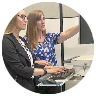
We work with project managers to create a reliable, versatile working space that can accommodate access to new equipment. Multiple users need to be taken in account, and how we can best enhance their workflow.
We offer a series of mounting arms which enables computers and medical devices to move weightlessly through a range of motion that can accommodate all users, whether they are sitting or standing. In addition to their counterbalanced vertical movement, the range also can be laterally adjusted, tilted, and swivelled to ensure devices are positioned at the optimal viewing angle.
Durability
All our Mounting Solutions are built with medical-grade materials and undergo rigorous safety and reliability testing to ensure they withstand the expected use of a Pathology environment.
Cleanability
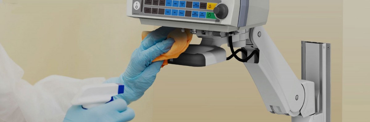
The Pathology environment requires solutions which can be cleaned frequently. Project Managers can have peace of mind that our Mounting Solutions undergo chemical compatibility testing with common cleaning agents used in the Pathology.
Service, and safe installation
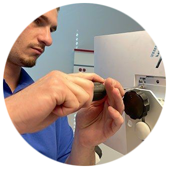
With new equipment, and evolving equipment – it’s important to make sure you have technical support, on-site services and engineering staff at your every call. Working with you every step of the way, from demos and procurement to installations and ongoing maintenance.
Our maintenance staff are fully aware of all risks associated with work in the laboratory and with laboratory equipment. Appreciating that much of the work in a clinical laboratory is concerned with handling specimens and materials that are infectious. Parity staff are experienced handling equipment which has been used to process those materials. Ensuring that equipment is decontaminated and cleaned, and know that they may require a certificate of decontamination before starting work.
From Wall Mounts, to Medical Keyboards/Mice and Monitors/All-In-One PCs
Pathology is a high contamination area, and all new equipment is often used by multiple users who can cross-contaminate if correct measures are not taken. The following extract is taken from cleaning compliances in a clinical laboratory:
“…fittings and furniture also need to be constructed from hard-wearing, easily cleaned materials with impervious surfaces that are resistant to damage by stains or chemicals.”
https://www.hse.gov.uk/pubns/clinical-laboratories.pdf
Mounting Solutions:
With infection prevention as important as ever, GCX solutions are manufactured under ISO 13485-certified quality standards and made with medical-grade materials that hold up to rigorous sanitation protocols and harsh cleaning agents. In addition to conducting wipe tests with a wide range of cleaning agents, GCX designs and constructs parts and equipment for easy, thorough cleaning throughout the day. This includes easily removed covers and unibody construction on many products to prevent germs from lingering in hard-to-reach crevices.
When it comes to product durability and cleanability, consider:
• All Wall Mount surfaces undergo wipe tests with cleaning agents ranging from various concentrations of isopropyl alcohol and Sani-wipes to commercial products like Lysol and Windex.
• Our easily removed channel covers help hide cables and make cleaning quick and stress-free.
• GCX arms like the VHM-T and VHM-P have a seamless, unibody construction that prevents debris and germs from collecting in hard-to-reach crevices.
• Materials are medical-grade to withstand constant usage and hospital-level cleaning regimens; whereas, commercial solutions typically are created for less rigorous, infrequent use.
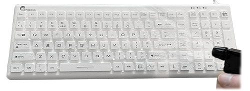
Medical keyboards and mice
The following extract is taken from cleaning protocols in a clinical laboratory:
“Staff should avoid contaminating bench surfaces and equipment. While gloves may adequately protect the wearer, care is needed to prevent contamination of equipment such as telephone handsets and computer keyboards. Plastic covers resistant to repeated treatment with disinfectant can protect equipment, such as keyboards, from contamination.”
https://www.hse.gov.uk/pubns/clinical-laboratories.pdf
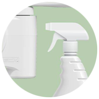
We provide cleanable keyboards and mice designed to withstand frequent disinfection. Offering the highest cleaning compliance*, and perfect for Pathology areas where the risk of contamination is high.
*They are IP68 which means the keyboards are dust-tight and protected against complete, continuous immersion in water.
Medical All-In-One PCs and cleanable Displays
Each Pathology department features many displays/screens for viewing information access. It’s essential that those displays are rugged, with longevity to be cleaned repeatedly – they need to be designed for healthcare environments. In the past, we have seen commercial displays being installed, which are not fit for purpose, and cleaning would damage the technology.
The Computers and displays we provide are built for 24/7 Hospital environments, and can be frequently disinfected. Often, we will offer a demonstration to showcase various solutions to suit different workflow. All of which are easy to clean and laboratory compliant.
We provide various scanners and printers to provide complete workflow solutions.
For example, Zebra has developed a labelling solution for histology labs to easily and reliably identify tissue specimen slides. Labs process thousands of samples each year, enabling diagnosis of a variety of diseases. From patient to slide creation, the correct identification of tissue is vital. A study of errors in histology in a U.S. hospital* showed that 35% of errors were detected in the labelling stage of sample processing. It was found that the introduction of technology and the adoption of automatic tracking systems dramatically reduced errors in specimen processing.
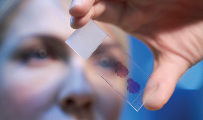
Staining slides
Cells are colourless, so to enable examination and diagnosis, staining techniques are essential to make the tissue visible down the microscope. The mostly widely used staining technique used in histopathology is haematoxylin and eosin (H&E) in which the nuclei of the cells are stained blue and the cytoplasm and other tissues are stained various shades of pink. This shows the cellular changes associated with disease.
Specially designed to resist the dip cycles of xylene and graded alcohols involved in this process, yet remain legible and scannable, Zebra’s 8000T Lab Resist label material is ideal where standard labels would fail.
Printer, ribbon and scanner:
By providing the printer, ribbon and scanner to create durable, high-quality labels to improve process flow and maintain a link between patient and sample. With Zebra’s easy-to-use ZD420 cartridge printer, users can quickly and efficiently print labels on-demand.
In order to follow best practice, a sample should be labelled as early as possible and given an identity at the start of the slide creation process.

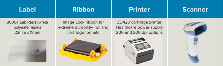
Our printer/scanner range is designed for easy cleaning. For example, the Zebra printer is created with disinfectant-ready plastics and a sealed button interface that make it easy to clean and disinfect. And, the power supply is IEC 60601-1 compliant for use in healthcare facilities.
When providing printers, and scanners – we would incorporate the information stated earlier regarding workspace best practices. Ensuring they were mounted in a safe, compliant way that aided workflow.
Viewing software
After the file has been created, specialised software often compresses the image so it has an optimal size for viewing and analysis. After that, the digital slide can be viewed on a display.
Display
Using the viewing software, the whole slide image can be brought up on a display for analysis by the pathologist. They can use the display as a tool next to their microscope, but the market also offers whole slide imaging systems that were designed for digital pathology and received regulatory clearance to be used for primary diagnosis.
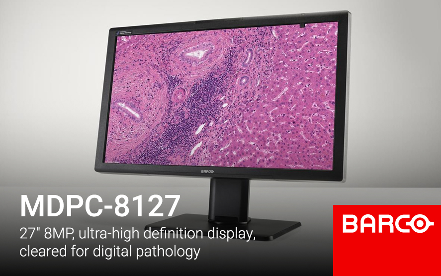
We offer stand-alone displays that can be used for primary diagnosis, and are a UK distributor of Barco.
Just like a microscope, the display is the device through which the pathologist views a specimen. Needless to say, it’s important that this device is reliable and shows the slide in an optimal and consistent way.
The Barco MDPC-8127, is a ultra-high definition medical grade display designed exclusively for digital pathology. With regulatory clearances for use in digital pathology including primary diagnosis, it’s the first display that you can confidently integrate into your digital pathology workflow with multiple whole slide imaging systems.* Work with superior imaging technology and analyse your histological samples confidently, with unprecedented visual richness.
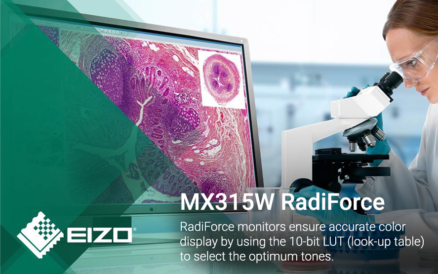
We also have options from EIZO with the EIZO RadiForce range also recommended. EIZO monitors are optimally equipped for this challenge. Not only do they have an excellent screen resolution, but they also display colours with outstanding accuracy. A high (spatial) resolution, a wide gamut, a deep black level, homogeneous illumination, a reproduction curve that can be calibrated according to individual requirements and consistent image reproduction quality guarantee reliable diagnostics, even for the smallest tissue structures.
Storage
Once a pathologist is done with a case, the specimen needs to be stored or archived. Just like the physical slide is stored in a dedicated space, digital files need to be stored in an information management system or vendor neutral archive. Laws vary across regions for how long this data needs to stay available.
Even though online storage of large quantities of digital slides brings along its own cost, gathering big data makes deep learning and the use of algorithms possible, the use of which can lead to better insights into specific diseases.
Digital Pathology is here, and has already started to transform most NHS Trusts.
Digital often means more technology, and that can mean creating more congested areas. We can help by clearing benches, and Mounting equipment – to help keep workspaces free. While also providing medical-grade equipment in-line with your cleaning protocols.
Get in touch to see how we can help you!
See our latest case studies, team updates and new products below. If you would like a case study at your hospital, please let the team know and we would be happy to feature it as well as do a video case study.
We’ve received your message and we will reply to your message as soon as possible.
If your enquiry is urgent, please contact us via phone or by live chat during our working hours for an immediate response.
